


Livestock Diseases
"Cara inchada" and cellular immunity in cattle
- Abstracts: English Portuguese
Abstract in English:
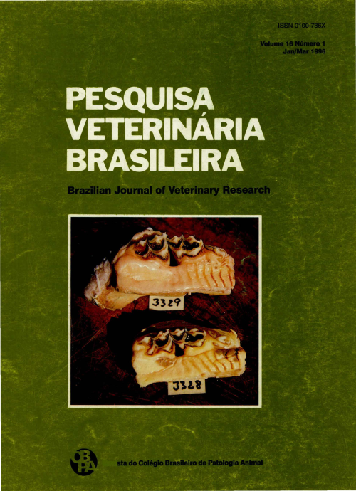
Attempts were made to study possible effects of the periodontal disease "cara inchada" (CI) on the cellular immunity of cattle, using adherence-, chemotaxis- and phagocytosis-determinations. Adherence of Bacteroides melaninogenicus to bovine granulocytes was significantly decreased in animals with CI. Phagacytosis of B. melaninogenicus by polymorphonuclear granulocytes (PMN) was also decreased in CI-diseased animals. Chemotaxis of the granulocytes appeared to be slightly increased in animals with CI and animals without CI coming from CI-affected herds.
Abstract in Portuguese:
Foram realizados estudos para verificar os possíveis efeitos da periodontite dos bezerros ("cara· inchada") sobre a imunidade celular, utilizando-se a determinação da aderência, fagocitose e quimiotaxia de Bacteroides melaninogenicus frente a granulócitos de animais com e sem lesões da enfermidade. A aderência de B. melaninoganicus foi significativamente inferior quando testada frente a granulócitos isolados de bezerros afetados pela "cara inchada" (CI). Da mesma forma, a fagocitose de B. melaninogenicuspor polirnorfonucleares (PMN) apresentou-se diminuida em animais com CI. Por outro lado, frente a B. melaninogenicus, o índice leucotático de PMN de bezerros com CI e de animais sadios do mesmo rebanho, pareceu levemente superior quando comparado com o de animais de área indene.
Experiments on the to:xicity of some ornamental plants in cattle
- Abstracts: English Portuguese
Abstract in English:
Due to inquiries about the toxicity of ornamental plants to farm animals, and the few data available in the literature on this subject, feeding experiments were performed in cattle with the following plants: Allamanda cathartica, Nerium oleander and Tbevetia peruvíana of the Apocinaceae family, Codiaeum sp., Euphorbia cotinifolia, Euphorbia pulcherrima and Euphorbia tirucalli of the Euphorbiaceae family, Datura arborea of the Solanaceae family, Colocasia antiquoruni, Dieffenbachia picta, Monstera deliciosa, Philodendron hastatum, Philodendron selloum ( = P. sellowii) and Scindapsus aureus ( = Epipremnum aureum) of the Araceae family, Rhododendron ledifolium and Rhododendron indicum of the Ericaceae family, and Malvaviscus arboreus of the Malvaceae family. The only plants which caused lethal poisoning were Allamanda cathartica, Neríum oleander, Tbevetia peruviana and Rhododendron indicum. The letal dose for A. catártica was 30 g/kg (causing death of 2 of 3 bovines which received this dose), for N. oleanderit varied from 0,25 to 0,5 g/kg (0,25 killed 1 of 4 and 0,5 the 2 bovines which received this dose); T. peruviana caused death of 3 bovines with doses of 14,4 g/kg or more (out of 7 bovines which received these doses), and R. indicum caused death of the animal which received the highest dose which could be given, this is 29,39 g/kg, whilst a second bovine which received 29 g/kg suffered only severe poisoning. Other plants that caused severe poisoning without lethal outcome were Euphorbia pulcherríma, in doses of 16,4 and 30 g/kg, Rhododendron ledifolium in one bovine which ingested 20 g/kg, the highest dose of this plant administered. Moderate poisoning was caused by Datura arborea in one bovine which ingested 40 g/kg, by Colocasia antiquorum in 2 bovines which ingested 4 and 5,9 g/kg and by Dieffenbachia picta in 2 bovines, which ingested 3, 7 and 4,8 g/kg of the respective plants. The other plants did not cause poisoning or only slight symptoms. Allamanda cathartica caused mainly manifestations of colic and the most importante post-mortem and histopathological findings were severe oedema of all parts of the wall of the rumen and reticulum, and congestion of the mucosa of the remaining digestive tract. Nerium oleander caused severe cardiac arrhythmia and severe diarrhoea, sometimes with blood. The post-mortem findings were generalized hemorrhages and the main histological lesion was necrosis of heart fibres. Tbevetia peruviana also caused heart arrhythmia and diarrhoea. Post-mortem and histopathological findings were negative with exception of one of the bovines which had diarrhoea, in which the contents of the rumen were putrid and those of the intestine were liquid. Euphorbia pulchenima caused severe weakness. Datura arborea caused the symptoms of atropine poisoning. Rhododendron spp. caused mainly regurgitation without or with vomits, sialorrhoea, manifestations of colic, diarrhoea and instability. Post-mortem and histopathological findings in the only bovine that died, were negative. The plants of the Araceae family caused sialorrhoea and sublingual and submandibular oedema. It is concluded, that the rarity of the poisoning in cattle under natural conditions by these plants is due to its low palatability and the fact that normally these plants are not available to bovines, and yet that most of these plants have to oe ingested in large amounts to cause poisoning. An exception is Nerium oleander, whose lethal dose is low. It is practically the only ornamental plant of those tested in this study, which according to the data in the literature has caused cases of poisoning in cattle.
Abstract in Portuguese:
Em virtude de consultas sobre a toxidez de algumas plantas ornamentais em relação aos animais de fazenda, e os escassos dados na literatura sobre esse assunto, foram efetuados experimentos em bovinos com as seguintes plantas: Allamanda cathartica ("alamanda"), Nerium oleander ("espirradeira") e Thevetia peruviana ("chapeu-de-Napoleão") da família Apocinaceaé, Codiaeum sp. ("croton"), Euphorbia cotinifolia ("aiapana", "maleiteira"), Euphorbia pulchemma ("bico-de-papagaio") e Euphorbia tirucalli ("a veloz") da família Euphorbiaceae, Datura arborea ("trombeteira") da família Solanaceae, Colocasia antiquorum ("orelha-de-elefante"), Dieffenbachia picta ("comigo-ninguém-pode"), Monstera deliciosa ("costela-de-Adão"), Philodendron hastatum ("filodendron"), Philodendron selloum (=P sellowii) ("bananade-macaco") e Scindapsus aureus ( = Epipremnum aureum) ("jibóia, "jibóia-dourada") da família Araceae, Rhododendron ledifolium e Rhododedron indicum ("azaléia", "rododendron") da família Ericaceae, Malvaviscus arboreus ("papoula", "graxa-de-estudante", "chupetinha") da família Malvaceae. Verificou-se que as únicas plantas que causaram intoxicação grave com êxito letal, foram Allamanda cathartica, Nerium oleander, Tbevetia peruviana e Rhododendron indicum. A dose letal para A. cathartica foi de 30 g/kg, que matou 2 dos 3 bovinos que a receberam nesta dose, para N oleander foi de 0,25 a 0,5 g/kg (0,25 matou 1 de 4 e 0,5 g/kg os 2 bovinos que a receberam nessas doses); Tpenwiana causou a morte de 3 bovinos em doses a partir de 14,4 g/kg, entre 7 bovinos que a receberam nessas doses, e R. indicum causou a morte do bovino que ingeriu a maior dose que se conseguiu administrar, isto é 29,39 g/kg, enquanto um segundo que recebeu 29 g/kg adoeceu gravemente. Causaram intoxicação grave ainda, sem êxito letal, Euphorbia pulchemma, em doses de 16,4 e 30 g/kg, e Rhododendron ledifolium em um bovino que ingeriu 20 g/kg, a maior dose administrada desta planta. Intoxicação de intensidade moderada foi causada por Datura arbórea em um bovino que ingeriu 40 g/kg, por Colocasia antiquorum em 2 bovinos que ingeriram 4 e 5,9 g/kg e por Dieffenbachia picta em 2 bovinos que ingeriram 3,7 e 4,8 g/kg das respectivas plantas. As outras plantas não causaram intoxicação ou somente leves sintomas. Allamanda cathartica causou cólica como principal manifestação clínica e os achados de necropsia e histopatológicos mais importantes foram edema acentuado de todas as camadas da parede do rúmen e do retículo, além de congestão da mucosa do restante do tubo digestivo. Nerium oleander causou grave arritmia cardíaca e ainda acentuada diarréia, às vezes com sangue. Os achados de necropsia foram hemorragias generalizadas e a principal alteração histopatológica foi necrose de fibras cardíacas. Tbevetia peruviana também causou arritmia cardíaca e diarréia. Não foram verificadas alterações à necropsia e nos exames histopatológicos, a não ser em um dos bovinos que teve diarréia, em que o conteúdo do rúmen estava com cheiro pútrido e o de todo intestino estava líquido. Euphorbia pulchemma causou grande debilidade. Datura arborea causou sintomas de intoxicação por atropina. Rhododendron spp. provocaram principalmente regurgitamento sem ou com vômito, sialorréia, cólica, diarreia e perturbações de equilíbrio. Os achados de necropsia e histopatológicos no único bovino que morreu foram negativos. As plantas da família Araceae causaram sialorréia e edemas sublingual e submandibular. Conclui-se, que a raridade de casos de intoxicação por essas plantas, sob condições naturais em bovinos, deve estar ligada a dois fatores: à sua baixa palatabilidade e à falta de acesso dos bovinos a essas plantas de uma maneira geral, ainda mais que da maioria delas quantidades elevadas têm que ser ingeridas para causar quadro de intoxicação. Exceção constitui Nerium oleander; da qual doses pequenas das folhas já são letais. É praticamente a única planta ornamental das testadas nesse trabalho, sobre a qual há registros na literatura de casos de intoxicação natural em bovinos.
Radiological study of hereditary lymphedema in Hereford cattle
- Abstracts: English Portuguese
Abstract in English:
A radiological study of the fore and hind limb lymphatic system was performed in seven calves with hereditary hypoplasia. Four healthy calves from an unrelated Hereford herd were used as a contrai group. Sixteen calves, without signs of disease, from an experimental affected herd were also studied to detect subclinical cases of lymphedema. After sedation and local anaesthesia a trian-guiar flap of skin was reflected over the lateral aspect of the metacaÍpus and metatarsus to expose the subcutaneous lymphatics, which were previously stained by methylene blue injected subcutaneously into the interdigital space. The contrast medium was injected into the stained lymph vessels and lymphographies were taken in the anatomical regions where the popliteal and prescapular lymph nades are located. The lymphangiograms obtained were used to determine the caliber of lymph vessels and the cranio-caudal and proximo-distal dimensions of the popliteal lymph nades. It was demonstrated that direct lymphography is a suitable method to study the peripheral lymphatic system in the hind limbs of cattle with hereditary lymphatic hypoplasia. The lesions were hypoplasia and/or aplasia of the peripheral lymphatic system, characterized by decreased number and enlargement or absence of peripheral lymph vessels and decreased size or absence of popliteal lymph nodes. In calves without clinical signs the peripheral lymphatic lesions which would allow to detect subclinical cases were not observed.
Abstract in Portuguese:
Hipoplasia linfática heriditária foi estudada através do exame radiológico do sistema linfático periférico de sete bovinos da raça Hereford com diagnóstico clínico da doença. Quatro bovinos sadios, da mesma raça, pertencentes a um rebanho livre da doença foram utilizados como controle. Dezesseis bovinos sem sinais clínicos, pertencente a um rebanho experimental, no qual a doença foi reproduzida, foram, também, estudados com o objetivo de detectar-se casos subclínicos da enfermidade. Após a sedação dos animais e anestesia local, foi feita uma incisão na pele, no terço médio da face lateral dos ossos metacarpo e metatarso para exposição dos vasos linfáticos, marcados previamente por azul de metileno. Nos vasos linfáticos foi injetado meio de contraste e foram tomadas radiografias nas regiões anatômicas onde estão localizados os linfonodos pré-escapulares e poplíteos. Nas radiografias eram medidos o calibre dos vasos linfáticos e as dimensões dorso-ventral e crâneo-caudal dos linfonodos. Este estudo demonstrou ser a linfografia direta eficiente para a avaliação do sistema linfático periférico dos membros posteriores de bovinos com hipoplasia linfática. As lesões observadas, de hipoplasia e/ou aplasia do sistema linfático periférico, caracterizaram-se por redução no número e aumento do diâmetro ou ausência de vasos linfáticos e diminuição do tamanho ou ausência dos linfonodos poplíteos. Nos animais que não apresentavam sinais clínicos não foram observadas alterações no sistema linfático periférico que permitissem a detecção de casos subclínicos da enfermidade.
Diagnosis of phosphorus deficiency in cattle by histologic and microradiographic examination of ribs
- Abstracts: English Portuguese
Abstract in English:
The objective of the present study was to show the efficiency of histologic and microradiographic techniques performed on rib bane samples as a mean of diagnosing the phosphorus status of cattle raised on phosphorus deficient pastures and receiving different mineral supplements. The rib samples were obtained from cattle of diferente ages which died from disease that was clinically and epidemiologically diagnosed as botulism. Ten out of 24 juvenile and adult animals studied exibited alterations of osteomalacia, while the others without those alterations served as controls. It is suggested that the method could be useful to improve the diagnosis of phosphorus deficiency of cattle in Brazil.
Abstract in Portuguese:
O objetivo do presente estudo foi de mostrar a eficácia de exames histológicos e microrradiográficos de costelas para avaliar o "status" de fósforo em bovinos criados em pastagens deficientes neste elemento e que receberam suplementos minerais diversos. As amostras de costelas foram coletadas de animais de diferentes idades, os quais morreram de doença com diagnóstico clínico e epidemiológico de botulismo. Dez dos 24 bovinos estudados mostraram alterações de osteomalácia, ao passo que os outros sem estas alterações serviram de controles. Sugere-se que o método poderia ser útil para aperfeiçoar o diagnóstico da deficiência de fósforo em bovinos no Brasil.
Influence of subclinical staphylococci mastitis on physical, chemical and cellular milk characteristics
- Abstracts: English Portuguese
Abstract in English:
One hundred twenty-six milk samples from 63 apparently healthy cows, but positive in the California Mastitis Test were submitted to determinations of pH, acidity, density, butter-fat, total solids, non-fat solids, cryoscopic point, caseine level, chloride level And polymorphonuclear leukocytes. Forty-one cows were infected by coagulase-positive Staphylococcus and 22 by coagulase-negative Staphylococcus. The results obtained in the milk sample analysis from the healthy quarters and infected by coagulase-positive Staphylococcus showed variations of all constituents investigated. However only the pH values (F=4.17*) and the polymorphonuclear leukocytes (F=ll.35**) showed significant differences. On the other hand, between the milk samples from healthy quarters and infected quarters by coagulase-negative Staphylococcus, only the polymorphonuclear leukocytes (F=16.29**) showed significant differences.
Abstract in Portuguese:
Foram submetidas às determinações do pH, acidez titulável, densidade, teor de gordura, extrato seco total, extrato seco desengordurado, ponto crioscópico, teor de cloreto, teor de caseína e às contagens de leucócitos polimorfonucleares, 126 amostras de leite procedentes de 63 vacas aparentemente sadias, porém, positivas ao Califórnia Mastitis Test, das quais 41 mostraram-se infectadas por cepas de Staphylococcus coagulase positiva e 22 por cepas de Staphylococcus coagulase negativa. Apesar de ter sido observada a variação de todos os constituintes investigados entre as amostras de leite procedentes de quartos sadios e infectados por Staphylococcus coagulase positiva, apenas os valores de pH (F=4,17*) e das contagens de leucócitos polimorfonucleares (F=ll,35**), mostraram diferenças estatisticamente significativas. Por outro lado, verificou-se, também, que entre as amostras de leite oriundas de quartos sadios e infectados por Staphylococcus coagulase negativa, apenas os valores das contagens de leucócitos polimorfonucleares (F=l6,29**) apresentaram diferenças estatisticamente significativas.
Cross-reactions between Yersinia enterocolitica serotype 9 and Brucella spp in bovine and swine sera, in the area of Rio de Janeiro
- Abstracts: English Portuguese
Abstract in English:
The extent of antigen interference between Yersinia enterocolitica 0:9 and Bmcella spp was evaluated in 245 bovine serum samples divided into groups according to status of immunization against brucellosis, and in 119 swine serum samples. The specimens were submitted to the plate serum agglutination test, tube serum agglutination test and to the Rose Bengal test for Brucella spp, and to tube sérum agglutination for Y. enterocolitica 0:9. The immune response to Y. enterocolitica was demonstrated and considered significant in terms of its possible effects on the interpretation of serological tests for brucellosis. Agglutinating titers were dose or even similar from a quantitative viewpoint. Since in most cases it was not possible to determine precisely the etiologic agent, it is clearly necessary to set up a laboratory method for the diagnosis of brucellosis that will permit the distinction of specific infections.
Abstract in Portuguese:
O grau de interferência antigênica entre Yersinia enterocolitica 0:9 e Bmcella spp foi analisado em 245 soros de bovinos discriminados quanto ao estado de imunização contra brucelpse, e 119 soros de suínos. Os espécimens foram submetidos às provas de soroaglutinação rápida, soro-aglutinação lenta e antígeno acidificado para Brucella spp e soro-aglutinação lenta para Y. enterocolitica 0:9. A resposta imune a Y. enterocolitica foi evidenciada e considerada significativa em relação aos seus possíveis efeitos na interpretação dos testes sorológicos para brucelose. Os títulos aglutinantes foram aproximados ou mesmo similares do ponto de vista quantitativo. Como não foi possível determinar com precisão, na maioria dos casos, o agente etiológico, é necessário instituir um método laboratorial de diagnóstico de brucelose que permita distinguir as infecções específicas.
