 |
|
|
|
Year 2014 - Volume 34, Number 12
|

|
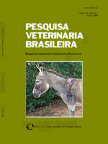
Outbreaks of primary photosensitization in equidae caused by Froelichia humboldtiana, 34(12):1191-1195
|
ABSTRACT. Knupp S.N.R., Borburema C.C., Oliveira Neto T.S., Medeiros R., Knupp L.S., Riet-Correa F. & Lucena R.B. 2014. [Outbreaks of primary photosensitization in equidae caused by Froelichia humboldtiana.] Surtos de fotossensibilização primária em equídeos causados por Froelichia humboldtiana. Pesquisa Veterinária Brasileira 34(12):1191-1195. Programa de Pós-Graduação em Medicina Veterinária, Universidade Federal de Campina Grande, Centro de Saúde e Tecnologia Rural, Campus de Patos, Av. Universitária s/n, Bairro Santa Cecília, Cx. Postal 61, Patos, PB 58708-110, Brazil. E-mail: sheilanribeiro@hotmail.com
The study was conducted in order to report outbreaks of photosensitization caused by Froelichia humboldtiana in equidae in the state of Rio Grande do Norte, Brazil. Animals from three farms and donkeys found abandoned in roads were examined. Peripheral blood samples were collected from five donkeys and two horses for analysis of serum activities of liver enzymes and concentrations of total, direct and indirect bilirubin. Skin biopsies were collected from two donkeys and a horse for histopatological exams. Fifty donkeys, 18 horses and two mules were affected. Of the affected donkeys, 45 were raised on roadsides and 30 died due to myiasis and weakness. The animals had a history of presenting skin lesions of photodermatitis about one month after being grazing in areas invaded by F. humboldtiana and recovered 10-30 days after being removed from these areas. If the animals were reintroduced in the paddocks with F. humboldtiana, pruritus and self-mutilation returned in a week or two. Young and adults donkeys showed extensive ulcerated wounds, that drained abundant serous exudate. All these wounds resulted from trauma caused by secondary self-mutilation to intense itching. In addition, many wounds had myiasis. The horses and mules had lesions of photodermatitis only in the areas of depigmented skin, no ocular lesions were observed. Histopathology of skin biopsies revealed perivascular inflammation in the superficial dermis. The epidermis had extensive ulcers, covered by fibrin associated with neutrophilic infiltrate and numerous basophilic bacterial aggregates. The serum activities of AST, GGT and serum concentrations of bilirubin were within normal ranges. The diagnosis of primary photosensitization associated with ingestion of F. humboldtiana was based on the epidemiology, clinical signs, serum biochemistry, skin lesions, and the recurrence of lesions after the reintroduction of the animals into the pasture invaded by the plant. F. humboldtiana is an important cause of primary photosensitization in equidae in the Brazilian semiarid region, resulting in weakness and death of large numbers of animals, mainly donkeys that don’t receive proper treatment. |
| |
|
|
| |
|
 |